The obex is also a.
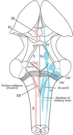
Cranial nerve nuclei present in floor of 4th ventricle.
10 3 is formed by the pons and medulla fig.
However the floor is the most related part to the cranial nerve nuclei.
The caudal tip of the fourth ventricle where it becomes the central canal is known as the obex.
Where is the nuclei for the third cranial nerve the oculomotor.
A cranial nerve nucleus is a collection of neurons gray matter in the brain stem that is associated with one or more cranial nerves.
A arrangement of the general afferent and efferent cell columns in the embryonic spinal cord.
The floor of the fourth ventricle the rhomboid fossa see fig.
Figure 12 1 arrangement of cranial nerve nuclei in the brainstem.
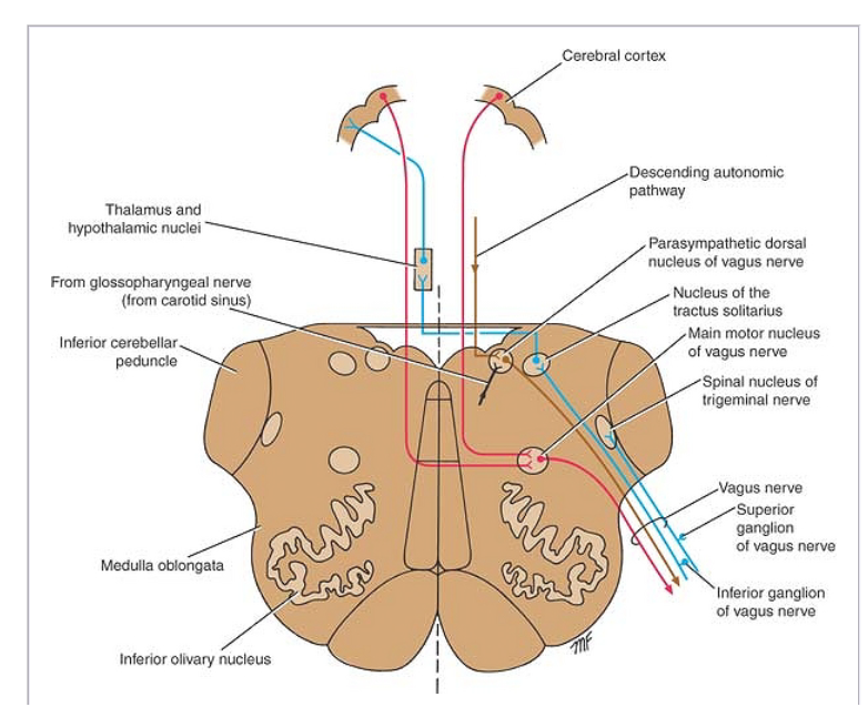
Surgical treatment of brainstem lesions carries a substantial risk of postoperative morbidity because of the risk of injuring the tightly packed cranial nerve nuclei cnn and neural tracts within the rhomboid fossa and brainstem lang et al 1991 historically neurosurgeons considered this area to be a no man s land with most lesions being inoperable baker 1965.
The 4th ventricle is a tent like cavity of the hindbrain lined with ependyma and filled up with cerebrospinal fluid csf it s situated in the posterior cranial fossa in front of the cerebellum and behind the pons and the upper part of medulla oblongata the cavity of the ventricle seems rhomboidal lozenge shaped in the horizontal section and presents a triangular outline in the sagittal.
We present two cases illustrating the benefit of utilizing intraoperative neurophysiological monitoring ionm for prevention of injuries to the lower cranial nerves during fourth ventricle tumor resection surgeries.
A lower triangular part formed by the upper part of the posterior surface of the medulla.
The floor of the fourth ventricle often called the rhomboid fossa because of its shape is divisible into an upper triangular part formed by the posterior surface of the pons.
Multiple cranial nerve nuclei are located on the floor of the fourth ventricle with a high risk of permanent damage.
The fourth ventricle has a roof at its upper posterior surface and a floor at its lower anterior surface and side walls formed by the cerebellar peduncles nerve bundles joining the structure on the posterior side of the ventricle to the structures on the anterior side.
At the level of superior colliculus in the midbrain.
Floor of the fourth ventricle below the facial colliculus facial nerve passes above it at the pons.
Axons carrying information to and from the cranial nerves form a synapse first at these nuclei lesions occurring at these nuclei can lead to effects resembling those seen by the severing of nerve s they are associated with.
B movement of these columns to the floor of the fourth ventricle in the embryonic rhombencephalon.